DJ-1
Protein-coding gene found in humans
| PARK7 | |||||||||||||||||||||||||||||||||||||||||||||||||||
|---|---|---|---|---|---|---|---|---|---|---|---|---|---|---|---|---|---|---|---|---|---|---|---|---|---|---|---|---|---|---|---|---|---|---|---|---|---|---|---|---|---|---|---|---|---|---|---|---|---|---|---|
 | |||||||||||||||||||||||||||||||||||||||||||||||||||
| Identifiers | |||||||||||||||||||||||||||||||||||||||||||||||||||
| Aliases | PARK7, DJ-1, DJ1, HEL-S-67p, Parkinsonism associated deglycase, GATD2 | ||||||||||||||||||||||||||||||||||||||||||||||||||
| External IDs | OMIM: 602533; MGI: 2135637; HomoloGene: 38295; GeneCards: PARK7; OMA:PARK7 - orthologs | ||||||||||||||||||||||||||||||||||||||||||||||||||
| |||||||||||||||||||||||||||||||||||||||||||||||||||
| |||||||||||||||||||||||||||||||||||||||||||||||||||
| |||||||||||||||||||||||||||||||||||||||||||||||||||
| |||||||||||||||||||||||||||||||||||||||||||||||||||
| |||||||||||||||||||||||||||||||||||||||||||||||||||
| Wikidata | |||||||||||||||||||||||||||||||||||||||||||||||||||
| |||||||||||||||||||||||||||||||||||||||||||||||||||


Protein deglycase DJ-1, also known as Parkinson disease protein 7, is a protein which in humans is encoded by the PARK7 gene.[5]
Structure
Gene
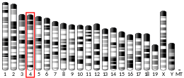
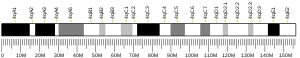
The gene PARK7, also known as DJ-1, encodes a protein of the peptidase C56 family. The human gene PARK7 has 8 exons and locates at chromosome band 1p36.23.[5]
Protein
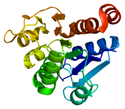
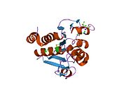
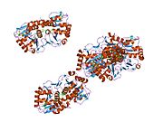
The human protein deglycase DJ-1 is 20 kDa in size and composed of 189 amino acids with seven β-strands and nine α-helices in total and is present as a dimer.[6][7][8] It belongs to the peptidase C56 family of proteins.
The protein structures of human protein DJ-1, Escherichia coli chaperone Hsp31, YhbO, and YajL and an Archaea protease are evolutionarily conserved.[9]
Function
Under an oxidative condition, protein deglycase DJ-1 inhibits the aggregation of α-synuclein via its chaperone activity,[10][11] thus functioning as a redox-sensitive chaperone and as a sensor for oxidative stress. Accordingly, DJ-1 apparently protects neurons against oxidative stress and cell death.[5] In parallel, protein DJ-1 acts as a positive regulator of androgen receptor-dependent transcription. DJ-1 is expressed in both the neural retina and retinal pigment epithelium of mammals, where it exerts a neuroprotective role against oxidative stress under both physiological and pathological conditions.[12][13]
Pyrroloquinoline quinone (PQQ) has been shown to reduce the self-oxidation of the DJ-1 protein, an early step in the onset of some forms of Parkinson's disease.[14]
Functional DJ-1 protein has been shown to bind metals and protect against metal-induced cytotoxicity from copper and mercury.[15]
DJ-1/PARK7 and its bacterial homologs: Hsp31, YhbO, and YajL can repair methylglyoxal and glyoxal glycated nucleotides.[16] Guanine, either in the form of a free nucleotide or as a nucleotide incorporated into nucleic acid (DNA or RNA), if glycated, can be repaired by DJ-1/PARK7.[16] Deglycase-deficient bacterial mutants with reduced ability to repair glycated bases in DNA show strong mutator phenotypes.[16]
DNA repair
DJ-1 is a DNA damage response protein that is recruited to sites of DNA damage where it participates in the repair of DNA double-strand breaks through the processes of non-homologous end joining and homologous recombination.[17] Evidence for a linkage between DNA damage and Parkinson's disease has been reported for decades.[17] Recently evidence has been presented that defective DNA repair is linked specifically to DJ-1 mutation, and thus DJ-1 mutation likely contributes to Parkinson's disease pathogenesis.[17]
Clinical significance
Defects in this gene are the cause of autosomal recessive early-onset Parkinson's disease 7.[5][18]
Interactions
PARK7 has been shown to interact with:
See also
References
- ^ a b c GRCh38: Ensembl release 89: ENSG00000116288 – Ensembl, May 2017
- ^ a b c GRCm38: Ensembl release 89: ENSMUSG00000028964 – Ensembl, May 2017
- ^ "Human PubMed Reference:". National Center for Biotechnology Information, U.S. National Library of Medicine.
- ^ "Mouse PubMed Reference:". National Center for Biotechnology Information, U.S. National Library of Medicine.
- ^ a b c d "Entrez Gene: PARK7".
- ^ "Uniprot: Q99497 - PARK7_HUMAN".
- ^ Honbou K, Suzuki NN, Horiuchi M, Niki T, Taira T, Ariga H, Inagaki F (Aug 2003). "The crystal structure of DJ-1, a protein related to male fertility and Parkinson's disease". The Journal of Biological Chemistry. 278 (33): 31380–4. doi:10.1074/jbc.M305878200. PMID 12796482.
- ^ Tao X, Tong L (Aug 2003). "Crystal structure of human DJ-1, a protein associated with early onset Parkinson's disease". The Journal of Biological Chemistry. 278 (33): 31372–9. doi:10.1074/jbc.M304221200. PMID 12761214.
- ^ Lee SJ, Kim SJ, Kim IK, Ko J, Jeong CS, Kim GH, Park C, Kang SO, Suh PG, Lee HS, Cha SS (Nov 2003). "Crystal structures of human DJ-1 and Escherichia coli Hsp31, which share an evolutionarily conserved domain". The Journal of Biological Chemistry. 278 (45): 44552–9. doi:10.1074/jbc.M304517200. PMID 12939276.
- ^ Shendelman S, Jonason A, Martinat C, Leete T, Abeliovich A (Nov 2004). "DJ-1 is a redox-dependent molecular chaperone that inhibits alpha-synuclein aggregate formation". PLOS Biology. 2 (11): e362. doi:10.1371/journal.pbio.0020362. PMC 521177. PMID 15502874.
- ^ Zhou W, Zhu M, Wilson MA, Petsko GA, Fink AL (Mar 2006). "The oxidation state of DJ-1 regulates its chaperone activity toward alpha-synuclein". Journal of Molecular Biology. 356 (4): 1036–48. doi:10.1016/j.jmb.2005.12.030. PMID 16403519.
- ^ Martín-Nieto J, Uribe ML, Esteve-Rudd J, Herrero MT, Campello L (August 2019). "A role for DJ-1 against oxidative stress in the mammalian retina". Neurosci Lett. 708: 134361. doi:10.1016/j.neulet.2019.134361. hdl:10045/94474. PMID 31276729. S2CID 195813073.
- ^ Shadrach KG, Rayborn ME, Hollyfield JG, Bonilha VL (2013). "DJ-1-dependent regulation of oxidative stress in the retinal pigment epithelium (RPE)". PLOS ONE. 8 (7): e67983. Bibcode:2013PLoSO...867983S. doi:10.1371/journal.pone.0067983. PMC 3699467. PMID 23844142.
- ^ Nunome K, Miyazaki S, Nakano M, Iguchi-Ariga S, Ariga H (Jul 2008). "Pyrroloquinoline quinone prevents oxidative stress-induced neuronal death probably through changes in oxidative status of DJ-1". Biological & Pharmaceutical Bulletin. 31 (7): 1321–6. doi:10.1248/bpb.31.1321. hdl:2115/53726. PMID 18591768.
- ^ Björkblom B, Adilbayeva A, Maple-Grødem J, Piston D, Ökvist M, Xu XM, Brede C, Larsen JP, Møller SG (2013). "Parkinson disease protein DJ-1 binds metals and protects against metal-induced cytotoxicity". Journal of Biological Chemistry. 288 (31): 22809–20. doi:10.1074/jbc.M113.482091. PMC 3829365. PMID 23792957.
- ^ a b c Richarme G, Liu C, Mihoub M, Abdallah J, Leger T, Joly N, Liebart JC, Jurkunas UV, Nadal M, Bouloc P, Dairou J, Lamouri A (July 2017). "Guanine glycation repair by DJ-1/Park7 and its bacterial homologs". Science. 357 (6347): 208–211. Bibcode:2017Sci...357..208R. doi:10.1126/science.aag1095. PMID 28596309.
- ^ a b c Wang ZX, Liu Y, Li YL, Wei Q, Lin RR, Kang R, Ruan Y, Lin ZH, Xue NJ, Zhang BR, Pu JL. Nuclear DJ-1 Regulates DNA Damage Repair via the Regulation of PARP1 Activity. Int J Mol Sci. 2023 May 12;24(10):8651. doi: 10.3390/ijms24108651. PMID 37239999; PMCID: PMC10218208
- ^ Bonifati V, Rizzu P, van Baren MJ, Schaap O, Breedveld GJ, Krieger E, Dekker MC, Squitieri F, Ibanez P, Joosse M, van Dongen JW, Vanacore N, van Swieten JC, Brice A, Meco G, van Duijn CM, Oostra BA, Heutink P (Jan 2003). "Mutations in the DJ-1 gene associated with autosomal recessive early-onset parkinsonism". Science. 299 (5604): 256–9. Bibcode:2003Sci...299..256B. doi:10.1126/science.1077209. PMID 12446870. S2CID 27186691.
- ^ Mukherjee K, Slawson JB, Christmann BL, Griffith LC (2014). "Neuron-specific protein interactions of Drosophila CASK-β are revealed by mass spectrometry". Frontiers in Molecular Neuroscience. 7: 58. doi:10.3389/fnmol.2014.00058. PMC 4075472. PMID 25071438.
- ^ Niki T, Takahashi-Niki K, Taira T, Iguchi-Ariga SM, Ariga H (Feb 2003). "DJBP: a novel DJ-1-binding protein, negatively regulates the androgen receptor by recruiting histone deacetylase complex, and DJ-1 antagonizes this inhibition by abrogation of this complex". Molecular Cancer Research. 1 (4): 247–61. PMID 12612053.
- ^ Takahashi K, Taira T, Niki T, Seino C, Iguchi-Ariga SM, Ariga H (Oct 2001). "DJ-1 positively regulates the androgen receptor by impairing the binding of PIASx alpha to the receptor". The Journal of Biological Chemistry. 276 (40): 37556–63. doi:10.1074/jbc.M101730200. PMID 11477070.
Further reading
- Cookson MR (Jan 2003). "Pathways to Parkinsonism". Neuron. 37 (1): 7–10. doi:10.1016/S0896-6273(02)01166-2. PMID 12526767. S2CID 14513509.
- Bonifati V, Oostra BA, Heutink P (Mar 2004). "Linking DJ-1 to neurodegeneration offers novel insights for understanding the pathogenesis of Parkinson's disease". Journal of Molecular Medicine. 82 (3): 163–74. doi:10.1007/s00109-003-0512-1. PMID 14712351. S2CID 32685319.
- Le W, Appel SH (Feb 2004). "Mutant genes responsible for Parkinson's disease". Current Opinion in Pharmacology. 4 (1): 79–84. doi:10.1016/j.coph.2003.09.005. PMID 15018843.
- Abou-Sleiman PM, Healy DG, Wood NW (Oct 2004). "Causes of Parkinson's disease: genetics of DJ-1". Cell and Tissue Research. 318 (1): 185–8. doi:10.1007/s00441-004-0922-6. PMID 15503154. S2CID 9453283.
- Pankratz N, Foroud T (Apr 2004). "Genetics of Parkinson disease". NeuroRx. 1 (2): 235–42. doi:10.1602/neurorx.1.2.235. PMC 534935. PMID 15717024.
- Heutink P (2006). "PINK-1 and DJ-1 — new genes for autosomal recessive Parkinson's disease". Parkinson's Disease and Related Disorders. Journal of Neural Transmission. Supplementa. Vol. 70. pp. 215–9. doi:10.1007/978-3-211-45295-0_33. ISBN 978-3-211-28927-3. PMID 17017532.
{{cite book}}:|journal=ignored (help) - Lev N, Roncevic D, Roncevich D, Ickowicz D, Melamed E, Offen D (2007). "Role of DJ-1 in Parkinson's disease". Journal of Molecular Neuroscience. 29 (3): 215–25. doi:10.1385/JMN:29:3:215. PMID 17085780. S2CID 85481215.
- Nagakubo D, Taira T, Kitaura H, Ikeda M, Tamai K, Iguchi-Ariga SM, Ariga H (Feb 1997). "DJ-1, a novel oncogene which transforms mouse NIH3T3 cells in cooperation with ras". Biochemical and Biophysical Research Communications. 231 (2): 509–13. doi:10.1006/bbrc.1997.6132. PMID 9070310.
- Taira T, Takahashi K, Kitagawa R, Iguchi-Ariga SM, Ariga H (Jan 2001). "Molecular cloning of human and mouse DJ-1 genes and identification of Sp1-dependent activation of the human DJ-1 promoter". Gene. 263 (1–2): 285–92. doi:10.1016/S0378-1119(00)00590-4. PMID 11223268.
- van Duijn CM, Dekker MC, Bonifati V, Galjaard RJ, Houwing-Duistermaat JJ, Snijders PJ, Testers L, Breedveld GJ, Horstink M, Sandkuijl LA, van Swieten JC, Oostra BA, Heutink P (Sep 2001). "Park7, a novel locus for autosomal recessive early-onset parkinsonism, on chromosome 1p36". American Journal of Human Genetics. 69 (3): 629–34. doi:10.1086/322996. PMC 1235491. PMID 11462174.
- Takahashi K, Taira T, Niki T, Seino C, Iguchi-Ariga SM, Ariga H (Oct 2001). "DJ-1 positively regulates the androgen receptor by impairing the binding of PIASx alpha to the receptor". The Journal of Biological Chemistry. 276 (40): 37556–63. doi:10.1074/jbc.M101730200. PMID 11477070.
- Bonifati V, Dekker MC, Vanacore N, Fabbrini G, Squitieri F, Marconi R, Antonini A, Brustenghi P, Dalla Libera A, De Mari M, Stocchi F, Montagna P, Gallai V, Rizzu P, van Swieten JC, Oostra B, van Duijn CM, Meco G, Heutink P (Sep 2002). "Autosomal recessive early onset parkinsonism is linked to three loci: PARK2, PARK6, and PARK7". Neurological Sciences. 23 (Suppl 2): S59-60. doi:10.1007/s100720200069. PMID 12548343. S2CID 13625056.
- Dekker M, Bonifati V, van Swieten J, Leenders N, Galjaard RJ, Snijders P, Horstink M, Heutink P, Oostra B, van Duijn C (Jul 2003). "Clinical features and neuroimaging of PARK7-linked parkinsonism". Movement Disorders. 18 (7): 751–7. doi:10.1002/mds.10422. PMID 12815653. S2CID 44253517.
- Miller DW, Ahmad R, Hague S, Baptista MJ, Canet-Aviles R, McLendon C, Carter DM, Zhu PP, Stadler J, Chandran J, Klinefelter GR, Blackstone C, Cookson MR (Sep 2003). "L166P mutant DJ-1, causative for recessive Parkinson's disease, is degraded through the ubiquitin-proteasome system". The Journal of Biological Chemistry. 278 (38): 36588–95. doi:10.1074/jbc.M304272200. PMID 12851414.
External links
- Overview of all the structural information available in the PDB for UniProt: Q99497 (Protein/nucleic acid deglycase DJ-1) at the PDBe-KB.
- v
- t
- e
PDB gallery
-
 1j42: Crystal Structure of Human DJ-1
 -
 1p5f: Crystal Structure of Human DJ-1
 -
 1pdv: Crystal structure of human DJ-1, P 31 2 1 space group
 -
 1pdw: Crystal structure of human DJ-1, P 1 21 1 space group
 -
 1pe0: Crystal structure of the K130R mutant of human DJ-1
 -
 1ps4: crystal structure of DJ-1
 - 1q2u: Crystal structure of DJ-1/RS and implication on familial Parkinson's disease
-
 1soa: Human DJ-1 with sulfinic acid
 -
 1ucf: The Crystal Structure of DJ-1, a Protein Related to Male Fertility and Parkinson's Disease
 -
 2or3: Pre-oxidation Complex of Human DJ-1


This article incorporates text from the United States National Library of Medicine, which is in the public domain.


